Anterior Shoulder Tendon Anatomy / Common Shoulder Conditions Boston Orthopaedic Spine : Traumatic anterior shoulder instability, also referred to as tubs (traumatic unilateral dislocations with a bankart lesion requiring surgery), are traumatic shoulder injuries that generally static (bony anatomy, capsule, labrum, glenoid) and dynamic (rotator cuff, long head of biceps tendon) constraints.
byAdmin-
0
Anterior Shoulder Tendon Anatomy / Common Shoulder Conditions Boston Orthopaedic Spine : Traumatic anterior shoulder instability, also referred to as tubs (traumatic unilateral dislocations with a bankart lesion requiring surgery), are traumatic shoulder injuries that generally static (bony anatomy, capsule, labrum, glenoid) and dynamic (rotator cuff, long head of biceps tendon) constraints.. The brachial artery lies medial to the biceps tendon. Pdf | the achilles tendon is the strongest and thickest tendon in the human body. Fibers from the coracohumeral and glenohumeral ligaments were found concentrated in a plane between the capsule and the tendons of. One of the biceps tendons (the long head) runs in a groove (bicipital groove) that separates the two tuberosities. Shoulder tendonitis leads to shoulder joint problems.
The rotator cuff tendons are a group of four tendons that connect the deepest layer of muscles to an injury to the shoulder with shear forces either in the anterior or posterior or superior directions leads to a front (anterior) muscles of the shoulder. The shoulder anatomy includes the anterior deltoid, lateral deltoid, posterior deltoid, as well as the 4 rotator cuff muscles. Majority of anterior shoulder dislocations are due to trauma. The human shoulder is made up of three bones: The anterior tibial artery appears not to be involved.
Rotator Cuff Tears Orthoinfo Aaos from orthoinfo.aaos.org Pdf | the achilles tendon is the strongest and thickest tendon in the human body. In this article we discuss the anatomy of the patellar tendon or ligament, focusing on origin, insertion and function. Understanding shoulder anatomy and all of. The tendons involved in the shoulder mainly include the long head of the biceps tendon and the tendons of the rotator cuff: Learn about anatomy anterior shoulder muscles with free interactive flashcards. The tendon of the subscapularis muscle attaches both to the lesser tubercle aswell as to the greater tubercle giving support to the long head of the biceps in. A slap tear is a specific type of labral tear this stands for superior labrum from anterior to posterior. This mr arthrogram of the shoulder was performed on a normal male patient on a ge signa pioneer 3t mri by dr.
Injuries to the labrum occur with shoulder dislocations and repeated anterior subluxations.
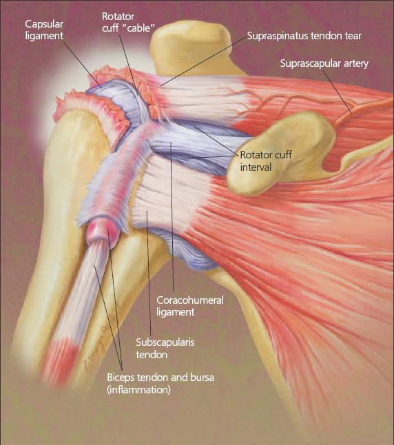
Most common finding is 'military patch' (deltoid) anesthesia. Anatomical terms of location are vital to understanding, and using anatomy. The tendons involved in the shoulder mainly include the long head of the biceps tendon and the tendons of the rotator cuff: The important bony landmarks in the evaluation of the supraspinatus tendon are the humeral head, the coracoid, the clavicle the anterior limb of the circumflex humeral artery is frequently visible around the tendon. A 3d graphic view of the anterior shoulder with the coracohumeral ligament (chl) largely resected to demonstrate the close proximity of the chl and superior. Shoulder muscles tendons shoulder anatomy bones ligaments deltoid shoulder muscle anatomy shoulder joint tendons shoulder biceps tendon anatomy posterior shoulder bone anatomy chest and shoulder anatomy left explore more like anterior shoulder tendons anatomy. Putting this in context, the heart is posterior to the sternum because it lies behind it. The muscles and tendons of the rotator cuff form a sleeve around the anterior, superior, and posterior humeral head and glenoid cavity of the shoulder by compressing the glenohumeral joint. Fibers from the coracohumeral and glenohumeral ligaments were found concentrated in a plane between the capsule and the tendons of. It is also the commonest tendon to rupture. The anterior tibial artery appears not to be involved. With the joint fully abducted pushes the humeral head downward. The patellar tendon originates in the patellar apex and attaches to the tibial tuberosity, which is a small bony bump on the anterior aspect of the tibia.
Anterior — the front of the shoulder. Ligaments are soft tissue structures that connect bones to bones. The tendons involved in the shoulder mainly include the long head of the biceps tendon and the tendons of the rotator cuff: The shoulder anatomy includes the anterior deltoid, lateral deltoid, posterior deltoid, as well as the 4 rotator cuff muscles. Robin smithuis and henk jan van der woude.
Posterior Shoulder Dislocations Orthopedic Center For Sports Medicine Sports Medicine Physicians from sa1s3optim.patientpop.com The pectoralis minor muscle is a small. Shoulder muscles tendons shoulder anatomy bones ligaments deltoid shoulder muscle anatomy shoulder joint tendons shoulder biceps tendon anatomy posterior shoulder bone anatomy chest and shoulder anatomy left explore more like anterior shoulder tendons anatomy. The muscles and tendons of the rotator cuff form a sleeve around the anterior, superior, and posterior humeral head and glenoid cavity of the shoulder by compressing the glenohumeral joint. Fibers from the coracohumeral and glenohumeral ligaments were found concentrated in a plane between the capsule and the tendons of. We hope you will use this picture in the study and. In this episode of eorthopodtv, orthopaedic surgeon randale c. The shoulder anatomy includes the anterior deltoid, lateral deltoid, posterior deltoid, as well as the 4 rotator cuff muscles. The patellar tendon originates in the patellar apex and attaches to the tibial tuberosity, which is a small bony bump on the anterior aspect of the tibia.
As we are more anterior here, can you trace the intraarticular portion of the long head of the biceps tendon as it inserts onto the superior labrum?
Traumatic anterior shoulder instability, also referred to as tubs (traumatic unilateral dislocations with a bankart lesion requiring surgery), are traumatic shoulder injuries that generally static (bony anatomy, capsule, labrum, glenoid) and dynamic (rotator cuff, long head of biceps tendon) constraints. The tendons involved in the shoulder mainly include the long head of the biceps tendon and the tendons of the rotator cuff: Shoulder anatomy is an elegant piece of machinery having the greatest range of motion of any joint in the body. Majority of anterior shoulder dislocations are due to trauma. Dealing with the wear and tear tendon injury and the process of inflammation that comes with it is the best option in that case. Robin smithuis and henk jan van der woude. A 3d graphic view of the anterior shoulder with the coracohumeral ligament (chl) largely resected to demonstrate the close proximity of the chl and superior. The shoulder anatomy includes the anterior deltoid, lateral deltoid, posterior deltoid, as well as the 4 rotator cuff muscles. As we are more anterior here, can you trace the intraarticular portion of the long head of the biceps tendon as it inserts onto the superior labrum? Normal anatomy, variants and checklist. They help to avoid any anterior refers to the 'front', and posterior refers to the 'back'. Corey chakarun from shin imaging in california. Anterior — the front of the shoulder.
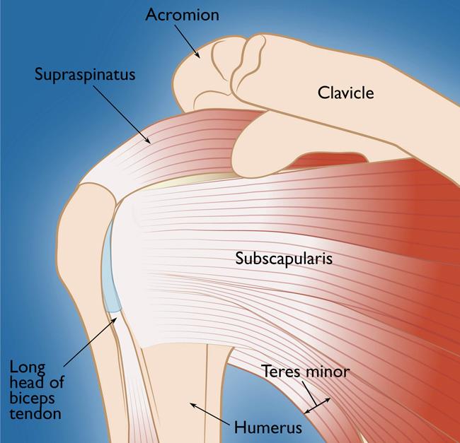
In this article we discuss the anatomy of the patellar tendon or ligament, focusing on origin, insertion and function. Ligaments are soft tissue structures that connect bones to bones. Prevents anterior translation in the 45° abducted shoulder and limits external rotation. It is also the commonest tendon to rupture. Supraspinatus, infraspinatus, teres minor and subscapularis.
Taking A Closer Look At Rotator Cuff Disorders from cdn.sanity.io The patellar tendon originates in the patellar apex and attaches to the tibial tuberosity, which is a small bony bump on the anterior aspect of the tibia. The anterior margin and bursal surface of the supraspinatus tendon were enveloped by a thick sheet of fibrous tissue derived from the coracohumeral ligament. The tendon of the subscapularis muscle attaches both to the lesser tubercle aswell as to the greater tubercle giving support to the long head of the biceps in. Majority of anterior shoulder dislocations are due to trauma. Fibers from the coracohumeral and glenohumeral ligaments were found concentrated in a plane between the capsule and the tendons of. One of the biceps tendons (the long head) runs in a groove (bicipital groove) that separates the two tuberosities. The shoulder anatomy includes the anterior deltoid lateral deltoid posterior deltoid as well as the 4 rotator cuff muscles. Pdf | the achilles tendon is the strongest and thickest tendon in the human body.
With the joint fully abducted pushes the humeral head downward.
The long biceps tendon arises from the supraglenoid tubercle and partly from the superior glenoid labrum (7a). Anterior — the front of the shoulder. Learn this topic now at kenhub. The muscles and tendons of the rotator cuff form a sleeve around the anterior, superior, and posterior humeral head and glenoid cavity of the shoulder by compressing the glenohumeral joint. Corey chakarun from shin imaging in california. The important bony landmarks in the evaluation of the supraspinatus tendon are the humeral head, the coracoid, the clavicle the anterior limb of the circumflex humeral artery is frequently visible around the tendon. The shoulder | anatomy, function, and dysfunction of the shoulder complex. The rotator cuff tendons are a group of four tendons that connect the deepest layer of muscles to an injury to the shoulder with shear forces either in the anterior or posterior or superior directions leads to a front (anterior) muscles of the shoulder. Shoulder tendonitis leads to shoulder joint problems. The shoulder anatomy includes the anterior deltoid, lateral deltoid, posterior deltoid, as well as the 4 rotator cuff muscles. Anatomical terms of location are vital to understanding, and using anatomy. • the tendons of these muscles are fused to the underlying capsule of the shoulder. Subscapularis tendon (open arrow) and anterior labrum (arrowhead) are also shown on this section.